X-Rays
Book Appointment
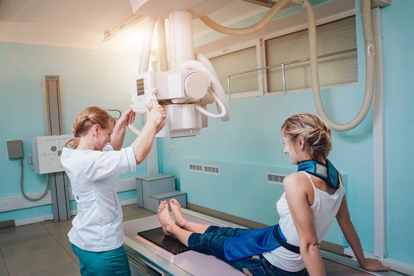
X-rays are a form of electromagnetic radiation, similar to visible light. Unlike light, however, x-rays have higher energy and can pass through most objects, including the body. Medical x-rays are used to generate images of tissues and structures inside the body. If x-rays traveling through the body also pass through an x-ray detector on the other side of the patient, an image will be formed that represents the “shadows” formed by the objects inside of the body
When are medical x-rays used?
When used appropriately, the diagnostic benefits of x-ray scans significantly outweigh the risks. X-ray scans can diagnose possibly life-threatening conditions such as blocked blood vessels, bone cancer, and infections. However, x-rays produce ionizing radiation—a form of radiation that has the potential to harm living tissue. This is a risk that increases with the number of exposures added up over the life of an individual. However, the risk of developing cancer from radiation exposure is generally small.
An x-ray in a pregnant woman poses no known risks to the baby if the area of the body being imaged isn’t the abdomen or pelvis. In general, if imaging of the abdomen and pelvis is needed, doctors prefer to use exams that do not use radiation, such as magnetic resonance imaging (MRI) or ultrasound. However, if neither of those can provide the answers needed, or there is an emergency or other time constraint, an x-ray may be an acceptable alternative imaging option.

